Mass spectrometry imaging
Mass spectrometry imaging (MSI) enables mapping the distribution of chemical species on various surfaces. Most often, the spatial distribution of lipids, peptides, proteins, drugs, and drug metabolites is visualized on tissue slices. The analytes are desorbed from the studied sample's surface, ionized, and introduced to a mass spectrometer. Mass spectra are collected from each point of the selected tissue area; point-to-point distance defines the image resolution. After that, the map of the selected ion can be constructed using specialized software. Various desorption/ionization techniques are used in MSI.
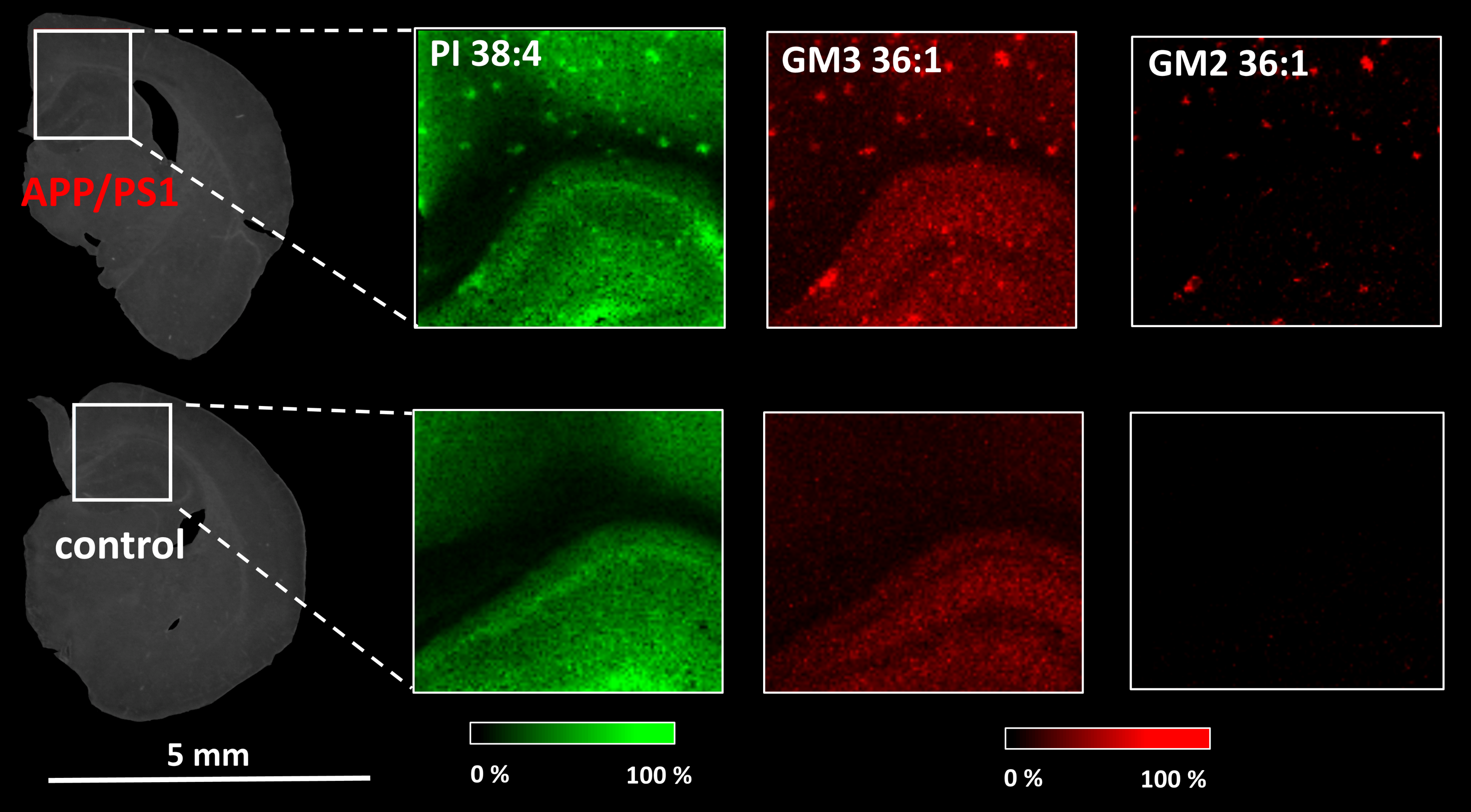
We have developed MSI based on matrix-assisted laser desorption/ionization (MALDI-MSI) to monitor lipids' spatial distribution within target tissues (mouse or rat brain, kidney, or liver). Various MALDI matrices were tested, and sample preparation procedures were optimized to achieve good selectivity for target lipid classes. Currently, we use MALDI-MSI to study neurodegenerative diseases, especially Alzheimer's disease (AD). AD is pathologically characterized by the accumulation of hyperphosphorylated tau neurofibrillary tangles and β-amyloid plaques in the brain. Recent evidence has shown that lipids are also involved in AD pathology. However, the exact underlying mechanism of AD is still not fully understood. Therefore, we use MALDI-MSI to monitor the spatial distribution of various lipids within target tissues to determine the lipid changes in mice models of neurodegeneration. In the cooperation with the group of Lenka Maletínská we also analyse the effect of potential neuroprotectives.